- Enabling the detection of global atmospheric variations over several hundred thousand years -
Satoshi Takeya, researcher, of Nano-Dynamics Analysis Research Group (Kazumasa Honda, Group Leader) of the Research Institute of Instrumentation Frontier (Shinngo Ichimura, Director) of the National Institute of Advanced Industrial Science and Technology (AIST; Hiroyuki Yoshikawa, President) jointly with Hitachi Ltd. (Kazuo Furukawa, President), Tsukuba University (Youichi Iwasaki, President), and the High Energy Accelerator Research Organization (KEK; Atsuto Suzuki, Director General), in collaboration with the Institute of Low Temperature Science, Hokkaido University (ILTS; Masaaki Wakatsuchi, Director), and the National Institute of Polar Research (NIPR), have succeeded in non-destructive 3D visualization of air-hydrate crystals, which transformed from ice and paleatmosphere confined in the ice, in Antarctic ice. This visualization has been achieved by developing a sample chamber for phase contrast X-ray CT device. The experimental method enables the detection of the global environmental variations which the earth has experienced for the past several hundred thousand years.
 |
|
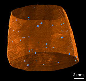
Air-hydrate grains distributed in an ice drilled from Antarctica
A side view of a see-through ice column (the outer surface is coloured orange) and air-hydrate crystalline grains (blue)
|
For addressing the issue of global warming, reliable data to verify various predictive models have been required. In particular, it is very important to clarify the correlation between variations in gas compositions, such as increases in CO2, and temperatures or properties of the atmosphere. Ice-sheets in the arctic and Antarctic regions preserve past atmosphere information, and thus the reconstruction of past climate and environmental variations based on the analysis of the data is expected.
In the arctic and Antarctic regions, snow settles without melting together with air. During the period of snowfall, air can be confined in ice as air bubbles by pressure due to the weight of the settled snow layer. At depth levels where the pressure exceeds 50 atm, air reacts with ice and gradually changes into air-hydrate crystals. Since the first report of air-hydrates in the ice of the arctic and Antarctic regions in 1982 (Nature), they have attracted attention as indices for the comprehending of climate and atmosphere variations which the earth has experienced. The air-hydrates preserve the air which existed hundreds of thousands of years ago.
The difference between the densities of air-hydrate crystalline grains and the surrounding ice is very small, conventional X-ray picture and X-ray CT methods cannot be applied for visualization of air-hydrates in ice. Thus, there have been no measuring methods for the 3D distributions of air-hydrates in ice or the density variations of the air-hydrates, while they are essential for understanding the formation mechanism of the hydrates and gas compositions in the hydrates.
Hitachi has developed a phase contrast X-ray CT method using an X-ray interferometer, and, jointly with Tsukuba University and KEK, has realized an imaging device enabling the non-destructive 3D observation of samples with a density resolution approximately 1000 times higher than that of conventional X-ray CT methods using synchrotron radiation (this device has been developed as the part of Special Coordination Funds of the Ministry of Education, Culture, Sports, Science and Technology). AIST has explored the applications of the phase contrast X-ray CT method, jointly with Hitachi, Tsukuba University, and KEK, and, has developed a low-temperature observation method for air-hydrates in ice since 2005, jointly with ILTS and NIPR.
The X-ray CT techniques widely used in medical care and industry construct 3D images by converting variations in transmittance (absorption) of X-ray passing through materials into image contrast. On the other hand, the phase contrast X-ray CT method constructs 3D images by converting phase variations (phase shifts) of X-rays passing through materials into image contrast. For samples composed of light elements (hydrogen, carbon, nitrogen, oxygen, etc.), the detection sensitivity is 1000 times higher than that of the conventional methods.
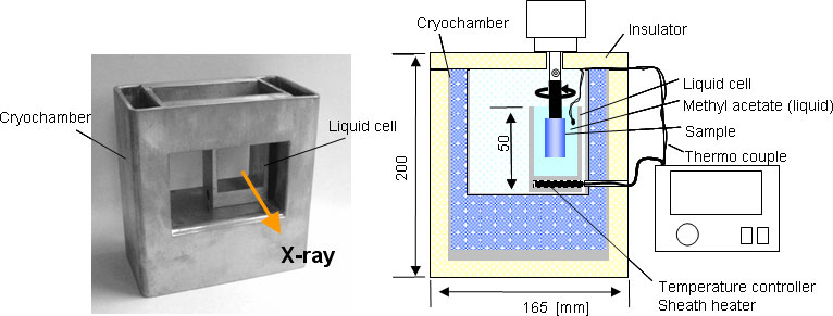
In this work, we have developed the sample chamber for the phase contrast X-ray CT device, and thereby enabled measurement at temperature range of -80°C to room temperature. The sample chamber enables the observation of samples of 15 mm in diameter and 20 mm in height, and its spatial resolution is 50 mm. Moreover, the phase contrast X-ray method enables not only 3D visualization of air-hydrates in ice but also the absolute density mapping with a density resolution of the order of mg/cm3.
Analytical results for south polar ice show that the density of air-hydrate crystalline grains is 937 mg/cm3, heavier by 14 mg/cm3 than that of the surrounding ice at -40°C. Furthermore, the observed density of the air-hydrates shows individual differences. It suggests that the deviation is caused by the crystallographic difference due to compositions and amounts of nitrogen, oxygen etc. included in the air-hydrates.
The phase contrast X-ray CT observation method, which enables a 3D display of the individual difference of the air-hydrate crystal densities, is a non-destructive and quantitative measuring method. The experimental technique will be a new significant method not only for the air-hydrate but also for various materials.
 |
|
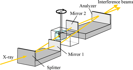
Measurement principle of a low-temperature phase contrast X-ray CT device
|
 |
|
Sample chamber
Left: Photograph of the sample chamber
Right: A set up diagram of the sample chamber in measurement time (an image viewed from the direction of incident X-ray beam)
|
The measuring time is approximately 30 min. currently. Hereafter, we plan to enhance the measuring efficiency of the device as an analysis tool by shortening the time. The improvement will lead to establish a system for systematic measuring of many samples of air-hydrate crystalline grains, and to advance the study of global environments by clarifying the history of climate and atmosphere variations. In addition, we plan to apply our technique to the measurement and non-destructive, in-situ observation of various functional materials in energy-related fields.