Update(MM/DD/YYYY):01/16/2018
Real Time Visualization with a Simple Device of Brown Adipose Tissues That Burn Fat
– Expected to accelerate development of therapy drugs for metabolic syndrome –
Presenters: Masako Yudasaka, Invited Senior Researcher, and Hiromichi Kataura, Prime Senior Researcher, of the Nanomaterials Research Institute, the National Institute of Advanced Industrial Science and Technology; Kumiko Saeki, Manager, Department of Infectious Diseases, Research Institute, National Center for Global Health and Medicine; Yuko Okamatsu-Ogura, Lecturer, of Hokkaido University; Kazuhiko Ishihara, Professor, of the University of Tokyo
Point
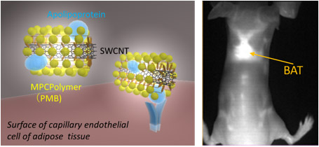
The researchers have developed a near-infrared fluorescent imaging method that uses single walled carbon nanotubes (SWCNTs) coated with PMB, a biocompatible MPC polymer. This is a probe that easily and affordably visualizes brown adipose tissues (BATs).
 |
|
Schematic drawing of deposition of PMB-coated SWCNTs on a BAT (left) and the imaging of the BAT between the shoulder blades of a mouse (right) |
New results
The researchers found that SWCNTs coated with the highly biocompatible dispersant PMB (PMB-SWCNTs) preferentially deposited in BATs when they were injected intravenously in mice. Irradiating the entire body of the mouse with a 730 nm wavelength LED and imaging with near-infrared light of wavelengths 1000 nm or longer fluorescent light from the PMB-SWCNTs made it possible to preferentially image BATs. In addition, since semiconductor SWCNTs that have high fluorescent intensities are separated and highly purified, the developed method is highly sensitive and can clearly image with a small amount of PMB-SWCNTs, which reduces the burden on a living body.
Background
A brown adipose tissue burns fat, but many aspects of this tissue remain unknown. In animal experiments on brown adipocytes and their activation, noninvasive observation is important for the purposes of preventing the suffering that animals endure in dissection and reducing the number of animals used. However, the only noninvasive observation method so far was the large and expensive PET-CT that uses radioactive isotopes. In contrast, a semiconductor SWCNT efficiently emits highly bio-permeable near-infrared light over 1000-nm wavelength allowing the clear fluorescence imaging of the mice vascular system. Until now there was no over 1000-nm fluorescence probe that can image living adipose tissues.
Future development
AIST will provide samples of PMB-SWCNTs with the aim of being utilized in animal experiments such as observing progress of changes in brown adipocytes that burn fat, for the purpose of research on prevention and treatment of metabolic syndrome.