Yumi Murata (Researcher) and Noriyuki Higo (Senior Researcher) of Systems Neuroscience Group, the Human Technology Research Institute (Director: Katsunori Matsuoka), the National Institute of Advanced Industrial Science and Technology (AIST; President: Ryoji Chubachi), and Takuya Hayashi (Unit Leader) and Hirotaka Onoe (Group Director) of Center for Life Science Technologies (Director: Yasuyoshi Watanabe), the Institute of Physical and Chemical Research (RIKEN; President: Ryoji Noyori), in collaboration with Yukio Nishimura (Associate Professor) and Tadashi Isa (Professor) of the National Institute for Physiological Sciences, the National Institutes of Natural Sciences, Takao Oishi (Associate Professor) of the Primate Research Institute, Kyoto University, and Hideo Tsukada (Manager), Central Research Laboratory, Hamamatsu Photonics K.K., revealed the changes in the brain that occur to take over motor functions that were lost due to brain lesion.
The researchers generated permanent lesions in the cerebral motor cortex in model animals and investigated changes in brain activity during the recovery process of motor function through rehabilitation. As a result, they discovered that the activity of the remaining brain areas, the ventral premotor cortex and the perilesional primary motor cortex, was changed and that those areas had taken over the function of the lesioned area. This result could be a key to developing neurorehabilitation, which is a new rehabilitative method based on the mechanisms of brain functions.
Details of the results will be published online in an American scientific journal, Journal of Neuroscience, on January 7, 2015 (Japan Time).
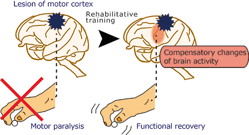 |
After the motor cortex that was responsible for motor command had been lesioned, rehabilitative training using a reach and retrieval task allowed recovery of the motor function of the hand.
Changes in brain activity, in which the remaining brain areas took over the function of the lost area, occurred during this process. |
Brain damage, including that caused by strokes, is becoming a serious social problem in Japan, whose society is aging. Because brain damage often leaves patients with sequelae, strokes are the leading cause of necessary nursing care after they occur. Impaired motor function of the hands, in particular, is a major factor that makes daily life inconvenient for patients. To reduce the burden on patients and their families, it is a very important task worldwide to make functional recovery through rehabilitation more efficient.
Recently, neurorehabilitation, a new rehabilitative method based on the mechanisms of brain recovery, has become the focus of attention. It is anticipated that it will be a promising method that will facilitate recovery better than conventional methods. However, the major problem has been that changes in the brain that achieve recovery were not well understood. The development of a more efficient rehabilitative method based on such changes would reduce the physical and financial burden on patients and their families, as well as the social burden of medical and nursing care. This would realize a society in which the elderly can live more actively.
AIST has strength in research on the mechanisms of brain recovery using model animals and has been involved in research and development projects on brain function recovery and the supporting technology. RIKEN has strength in measurement and analysis of brain activity. Using the strengths of both in this study, the researchers revealed the changes in brain activity that play an important role in the recovery of motor function in the hand.
This research was supported by the CREST program (FY2004-2009) and the PRESTO program (FY2009-2012) under the Strategic Basic Research Programs run by the Japan Science and Technology Agency, and Research Activity Start-up (FY2012-2013) and Scientific Research (C) (FY2013-2015) under Grants-in-Aid for Scientific Research run by the Japan Society for the Promotion of Science.
Using model animals, local lesions were induced in the area responsible for hand motor function in the primary motor cortex, which is central to providing motor commands from the cerebral cortex to the muscles. Hand motor paralysis occurred after the lesion, because the cerebral cortex was unable to provide a motor command. Movements requiring manual dexterity, such as holding something using the fingertips, are advanced motor functions that are shared only among humans and some animals. Because this function requires information processing by the cerebral cortex, recovery from lesion of the primary motor cortex was thought to be impossible. However, motor function of the hand, including movements requiring manual dexterity, was recovered approximately one month after the introduction of active rehabilitative training using a reach and retrieval task. It is thought that, during this functional recovery process through rehabilitative training, some changes occurred in the brain to compensate for the function of the lesioned primary motor cortex.
To capture these changes in the brain, positron emission tomography (PET) scans were used to assess brain activity during the performance of movements with manual dexterity. Before the lesion, activity was mainly observed in the primary motor cortex (Fig. 1 Left). When dexterous movements were recovered through rehabilitation after the lesion of the primary motor cortex, brain activity in the lesioned primary motor cortex was reduced compared with that before the lesion, whereas multiple brain regions showed more enhanced activity than that before the lesion. Just after the movements were recovered (early post-recovery stage), more enhanced activity was observed in another cortical region, called the ventral premotor cortex, compared with that before the lesion (Fig. 1 Right). Furthermore, during the plateau phase of recovery, which was several months after the lesion, changes in brain activity were observed in the primary motor cortex near the lesion (Fig. 2).
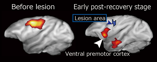 |
Figure 1: PET scan images before the brain lesion (left) and at the early stage of functional recovery (right)
The ventral premotor cortex was more active at the early post-recovery stage than it was before lesion. |
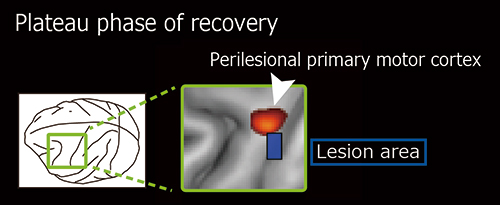 |
Figure 2: PET scan image during the plateau phase of recovery, which was several months after functional recovery
The perilesional primary motor cortex was active during the plateau phase after recovery. |
Clinical studies previously demonstrated that patients who have had strokes have altered brain activity compared with healthy people. However, the roles for each region of the brain were not identified, because it was thought that not all of these changes in brain activity were linked to functional recovery and that some changes might actually inhibit normal brain activity.
To investigate whether the changes in brain activity observed in this study might contribute to functional recovery, a drug, muscimol, was used to block the activity of the ventral premotor cortex and the perilesional primary motor cortex in the early post-recovery stage and the plateau phase of recovery. Blocking the activity of either area resulted in the recurrent impairment of hand movements, which confirmed that changes in the activity of these brain areas had taken over the area responsible for motor function of the hands in the lesioned primary motor cortex. It is possible that a new pathway to convey motor commands was established in the ventral premotor cortex and the perilesional primary motor cortex to compensate for the function of the lost area during the recovery process of motor function through rehabilitation.
Some research using model animals has proposed that changes in neuronal networks occur to take over the function of the damaged regions. In those studies, however, only part of the brain was investigated or the model animals were under anesthesia. Therefore, it was unclear whether altered neuronal networks were indeed used. In the current study, however, the researchers analyzed whole-brain activity during the performance of a reach and retrieval task. As a result, they captured changes in brain activity in the remaining undamaged area during the recovery process of motor function, and demonstrated that the changes in brain activity were essential for recovery. These findings are expected to directly lead to the development of rehabilitative technology.
AIST will analyze changes in gene expression caused by rehabilitation, whereas RIKEN will analyze changes in the neuronal network structure. Through these analyses, the detailed process of how rehabilitation produces changes in brain activity will be investigated from various perspectives. By providing these findings to medical technicians who are involved in rehabilitation, AIST and RIKEN will contribute to the development of new rehabilitative methods, electrical brain stimulation therapy that changes brain activity more directly, drugs that facilitate rehabilitation, and methods to assess rehabilitation effects.