- Dramatic sensitivity enhancement of antibody-based protein detection by integration of fluorescent quantum dots -
The National Institute of Advanced Industrial Science and Technology (AIST; Hiroyuki Yoshikawa, President) has succeeded in the synthesis of unique high quality fluorescent quantum dots, and the development of nano-biohybrid materials for protein and DNA/RNA measurements using quantum dots. Furthermore, we have made the first confirmation that the materials can be applied for detection of trace amounts of proteins in cell lysates using antibodies (immunoblotting).
The quantum dots are particles of several nanometers (one nanometer = 10-9 m) in diameter, which are formed by inorganic semiconductor substances. Because the quantum dots can emit strong fluorescence by ultraviolet irradiation, they have attracted attention for use as optical materials for bioimaging, biosensing, photosensitizing, etc., thus resulting in intense world-wide competition in their research and development. The AIST has succeeded in the preparation of high quality quantum dots which have the following characteristics: 1) high-luminescence performance, 2) effective size-distribution, 3) high photochemical stability, 4) non-aggregation, and 5) non-blinking. Furthermore, the AIST has successfully synthesized avidin- or biotin-conjugated quantum dots, and by the combination of both quantum dot hybrid materials, dramatically enhanced the sensitivity of antibody-based detection of proteins.
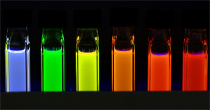 |
Fluorescence emitted from quantum dots.
Blue fluorescence can be emitted from small particles of approximately 2 nm in diameter, green from ~3 nm particles, yellow from ~4 nm particles, and red from large particles of ~5 nm.
The wavelength of the excitation light is 365 nm.
|
Hereafter, we will utilize this technique for the development of electrophoresis tips to detect and identify native proteins. Furthermore, we plan to develop surface treatment techniques for quantum dots to specifically label objectionable fungi and foreign substances invading various food production processes including the production of fermented food, and apply the techniques developed to evaluation of the optimal process management.
The results in this work have been published in
Journal of American Chemical Society, 2005,
127(26), 9328, and
Analytical Chemistry, 2006,
87(1), 321, which are both journals of the American Chemical Society. The results were also presented at "NanoTech 2006 International Nanotechnology Exhibition & Conference" held at Tokyo Big Sight on February 21-23.
For the past 10-15 years, the companies producing organic fluorescent dyes have recommended avidin-conjugated organic fluorescence dyes for the fluorescence detection of proteins with the immunoblot method. However, this method has not been used widely because of the following disadvantages of organic fluorescent dyes.
-
Organic fluorescent dyes soon experience photo-bleaching. Consequently, their fluorescent signals change, making quantitative measurements difficult.
-
When many organic fluorescent dyes are locally concentrated, self-quenching of fluorescent signals may be induced, resulting in drastic reduction in the signals.
-
Usually, proteins are labeled by one molecule of an organic fluorescent dye, leading to a considerable restriction of detection sensitivity.
For these reasons, improvement of the immunoblot method has been desired. Thus, our attention has been drawn to quantum dots, because they are not only fluorescent, but also nano-sized particles. When the quantum dots are covered by various inorganic or organic materials, they do not exhibit fluorescence self-quenching, even if they are highly concentrated. Also, we considered that, as biotin is able to strongly bond to avidin, if biotin-conjugated quantum dots and avidin-conjugated quantum dots are synthesized, many quantum dots can be integrated in a sandwich type. Thus, many quantum dots can be integrated in a local region of target proteins, and thereby fluorescent signals from target proteins can be intensified.
The quantum dots we have synthesized are composed of CdSe, which are particles with a diameter of 3-5.5 nm (1 nm = 10-9 m) as shown in Figure 1. When ultraviolet light is radiated onto the quantum dots, they can emit a brilliant fluorescence of blue to red, depending on particle size. These quantum dots exhibit sharp fluorescence spectra (Figure 2), thus being favorable for bio-imaging etc.
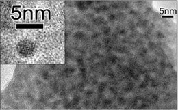
Figure 1 A high-resolution transmission electron microscope image of the quantum dots of 3 nm in diameter we have synthesized |
|
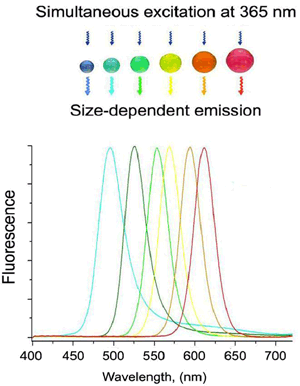
Figure 2 Fluorescence spectra depending on the size of quantum dots |
|
|
We have developed a technique for enabling the conjugation of various biomolecules to quantum dots, and, using this technique, we have synthesized avidin-conjugated and biotin-conjugated quantum dots (Figure 3).
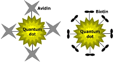 |
Figure 3 Schematic illustrations of avidin-conjugated and biotin-conjugated quantum dots.
Left: avidin-conjugated quantum dot, Right: biotin-conjugated quantum dot. |
Furthermore, we have applied these materials for detection of proteins in trace amounts using immunoblotting technology.
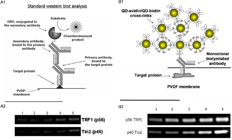 |
Figure 4 Detection of trace amounts of proteins (TRF1 and Tin2) existing in the cell.
Left, The conventional immunoblot method: A1, Scheme; A2; Bands of TRF1 and Tin2.
Right, The conventional immunoblot method with quantum dots: B1, Scheme; B2; Bands of TRF1 and Tin2.
The numbers indicate protein concentrations of cell lysate applied to each gel patch.
1: 10 µg 2: 20 µg 3: 30 µg 4: 40 µg 5: 50 µg.
TRF1: Telomeric binding factor (56 KDa)
Tin2: TRF1-interacting nuclear protein 2 (40 kDa)
Both are trace amounts of proteins existing in chronic myelogenous leukemia cell. |
As shown in Figure 4B1, the principle is as follows. First, an antibody specifically recognizes the target protein and binds to it. Next, a quantum dot-avidin bonds to the biotin-labeled antibody. Then, a quantum dot-biotin bonds to the quantum dot-avidin. Furthermore, a quantum dot-avidin bonds to the quantum dot-biotin. By repeating these steps, many quantum dots are accumulated one after another on one antibody, resulting in an increase in the intensity of fluorescence. Figure 4 shows schematic diagrams for the principle of the conventional immunoblot method (A1, chemiluminescence detection) and the new immunoblot method with quantum dots we have developed in this work (B1, fluorescence detection), respectively.
Using this method, we have detected two kinds of proteins, Telomeric Binding Factor (TRF1, 56 KDa) and TRF1-interacting nuclear protein-2 (Tin2), which are related to cell proliferation. These proteins are generated in chronic myelogenous leukemia cells of more than 90%, but the amount is very small. For comparison, we also have executed the detection of the same proteins using the conventional immunoblot method utilizing chemiluminescence.
As a result, we have confirmed that the quantum dot-based immunoblot method (Figure 4B2) enables ultra-sensitive detection for trace amounts of proteins directly in cell lysates which are undetectable with the conventional immunoblot method (Figure 4A2).
In addition, we have confirmed that signals from the quantum dots are stable for 40 minutes, being suitable for the uptake and analysis of data. Also, by light shielding at 4 °C, fluorescent signals are barely changed and stable for several weeks.
Consequently, these results evidently demonstrate that the "sandwich" immunoblot method with quantum dots is superior to the conventional immunoblot method in detection of trace amounts of proteins.