The National Institute of Advanced Industrial Science and Technology (AIST), an independent administrative institution, (president: Prof. Hiroyuki Yoshikawa) has developed a technique for controlling cell adhesion to a substrate surface through an optical means, by use of innovative polymer of which interaction with cells varies on light illumination.
In the cutting edge medical care of the 21st century, the utilization of living human or animal cells is supposed to acquire increasingly greater significance. For using living cells in the medical care, it is essential to segregate and purify cells of a particular type, through flow cytometry, magnetic cell separation or other means. However, as these segregation techniques require complete dispersion of cells in a solution, scaffold-requiring cells, or most of animal cells would be damaged through these processes.
Under such a circumstance, the AIST has thought of an idea that selecting and segregating cultured scaffold-requiring cells in situ would solve the problems, and developed a substrate to change cell adhesion when illuminated with light, or “photo- responsive cell culture substrate (PRCCS)”. It is possible to augment the cell adhesion by illuminating cells cultured on PRCCS with light of specific wavelength, and to reduce it, on the contrary, by illuminating with light of different wavelength. The light illumination and associated ON-OFF of cell adhesion affects very little the cell viability.
With the newly developed technique, it will be made possible while observing cultured cells under a microscope to peel off and collect cells of particular type only, or to remove all the other cells while keeping aimed ones as they are. Though there have been from earlier a number of methods to segregate selected cells, none of them have been able to collect selectively cells or cell groups of particular category under observation. It is expected that the new technique will provide an innovative experimental tool in the area of cell segregation.
Selection and separation of adhering cells by cell shapes
The 21st century may be regarded to be the age of biotechnology. Above all, the medical technology using live cultured cells is expected to open the way to the applications to diseases and injuries having been regarded to be difficult to cure, such as recovery of dysfunctional organs and tissues and healing of cancers and autoimmune diseases. Most of available somatic cells from experimental animals have experienced a certain degree of differentiation, and require some scaffolds for expressing their original functions by culturing and proliferating on certain kinds of substrate. With the conventional technique based on dispersed culture, it has been inevitable to lose the original functionality owing to damages on the cell surface caused by the process of separation from the substrate. For this reason, a technique has been demanded to handle scaffold-requiring cells in situ as cultured on the substrate.
Under such a circumstance, the AIST has been claiming the need of establishing cell manipulation technique to manipulate selected cells and cell groups on the surface of substrate. It has been known from earlier to culture cells in conformity with a certain pattern having been created on the surface of substrate, for instance by Prof. Takehisa Matsuda, Faculty of Medical Sciences, Kyushu University. In this method, however, an area to facilitate cell adhesion has to be prepared beforehand on the substrate surface by such a means as lithography, and the pattern cannot be changed after starting the culture. Prof. Mitsuo Okano, Tokyo Women's University, has developed a method to collect cells cultured on a substrate without giving damage by changing the substrate temperatures, though with this technique it is not possible to separate and collect particular cells and cell groups.
The AIST has recognized the merits of light as a control means, such as remote manipulation, immediate action and localized effects, and hit on an idea that the preparation of cell culturing substrate with polymer materials with cell adhesion affected by illumination would make it possible to control adhesion and attachment/ detachment of particular cells or cell groups. Based on this idea, the AIST applied for the Fiscal 2002 Industrial Technology Research Promotion Program of the New Energy and Industrial Technology Development Organization (NEDO), another independent administrative organization, (President: Dr. Tsutomu Makino) and after having been accepted, the present research work has been initiated.
· Photo-Responsive Cell Culture Substrate
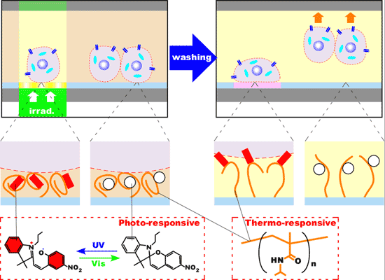
In order to attain this goal, the AIST has prepared innovative polymer by combining poly-N-isopropyl-acryl-amide, a thermo-sensitive polymer, with a photo- sensitive dyestuff of which structure may be affected by illumination with light of specific wavelength. The photopolymer has structure changed on light illumination at 37°C, cell-culturing temperature (Fig. 1). The dyestuff contained in the photopolymer acts upon the cell membrane to provide it with “capturing” role, making cells adhere to substrate. The affinity of dye to cell membrane is affected by the illumination, and it may be expected to drastically change the cell adhesion to the substrate on light illumination, if the latter surface is coated with the photopolymer.
 |
|
Fig. 1. A schematic diagram of cell adhesion control by use of photo-responsive polymer |
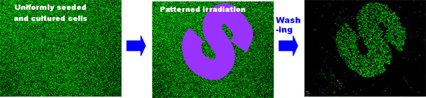
Under such an idea, a number of polymers have been synthesized, and the preparation of substrate surface has been studied in detail, resulting in a variety of cell patterning as shown in Fig. 2.
By the way, cells in non-illuminated area can be removed collectively by cooling, and it is possible to recover cell adhesion by illuminating vacant area. It is also possible to reduce cell adhesion by illuminating with light of different wavelength. That is, the cell adhesion in particular areas of substrate can be controlled on-demand basis.
 |
|
Fig. 2 Examples of cell separation using optical means |
|
After inoculation and culturing cells, an S-shaped pattern is illuminated with light of specific wavelength. On washing out the culture, cells in the S-pattern are kept intact, with individual living cells represented by green spots. |
· Application to 2D Cultured Cells Manipulating System
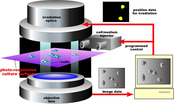
Cells cultured on the PRCCS can be manipulated through optical means in various ways. The AIST is planning to manufacture on a trial basis a general-purpose two-dimensional (2D) cultured cell manipulating system, as shown in Fig. 3.
 |
|
Fig. 3 A general-purpose 2D cultured cell manipulating system |
With this system, it may be possible either to remove or to keep intact cells of particular shape by selecting manually or by use of an image recognition program based on geometrical identification under a microscope, with selected sites illuminated, as shown in Fig. 4.
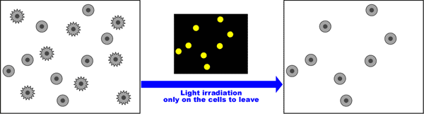
Most of available techniques of cell separation, such as flow cytometry and magnetic cell segregation, where antibodies specifically combining with target cells are utilized, are not applicable to cells of which antibodies are not known. However, as a number of cells have been found clearly identifiable by shape, such as primary culture cells and cancer cells, techniques for selecting and separating based on the shape recognition have been demanded. It is expected that the newly developed method may lead to the world first commercialization of cell selecting and separating devices based on shape recognition.
Fig. 4. Selection and separation of adhering cells based on cell shape
Besides, it is possible to switch the cell adhesion to the substrate repeatedly by illuminating the cell alternately with light of different wavelengths. As shown in Fig. 5, for instance, complicated patterning may be provided to cells after the inoculation and culture.

Fig. 5. Cell patterning after the culture