The Human Stress Signal Research Center (HSSRC) of the National Institute of Advanced Industrial Science and Technology (AIST), an independent administrative institution, has succeeded in developing screening technology to identify accurately environmental chemicals affecting the brain, in collaboration with the Endocrine Disruptors & Dioxin Research Project of the National Institute for Environmental Studies, another independent administrative institution.
Recently, the impacts to biological systems of some chemicals existing in the environment, such as resin-related chemicals, plasticizing agents, pesticides and so on are attracting attentions. Among them, certain substances are known to exercise disrupting effects to the endocrine function related to animal reproduction, and are called endocrine disruptors or environmental hormones. So far, studies have been focused on the effects of these chemicals to the genital organs of animals. In recent years, however, concerns are being extended to the possibility of certain environment chemicals causing mental disorders, and simple methods for identifying these hazardous factors out of environment chemicals are being sought for.
The newly developed screening technique consists in administering suspected environment chemicals to neonatal rats and evaluating changes in spontaneous motor activities. This simple method will make it possible to find out chemicals possibly responsible to mental disorder which were hardly detectable previously. Generally speaking, for assessing impacts of chemicals, the latter are administered to pregnant rats, and their effects to fetuses or young rats are studied. However, as the underlying mechanism is complicated by the involvement of changes in mother rats, it has been difficult to assess the action of chemicals to the brain.
The screening technique is effective enough for accurately identifying chemicals related to brain developmental disorders, such as pervasive developmental disorders (PDD) including autism, and attention deficit hyperactivity disorder (ADHD). The HSSRC-AIST and its partner have obtained hopeful findings on chemicals possible affecting the brain development through behavioral analyses and histological studies for two groups of rats: those directly administered with suspected chemicals, and model rats for brain developmental disorders, demonstrating the validity of this method.
The application of the newly developed technique is expected not only to be helpful for drafting the regulation for chemicals affecting the brain development, but also to contribute to the creation of better chemical substitutes and the development of new drugs effective for prevention or treatment of mental disorders.
In the modern life, we are benefited from various chemicals without doubt. Recently, however, adverse effects of environmental chemicals to the human body have come into question and a number of demerits have been revealed.
On the other hand, an increasing amount of attention is being paid to mental disorders. PDD including autism and ADHD are characterized by unfitting hyperactivity, impulsive insanity, carelessness, and communication failure, and in particular, hyperactivity disorders occur at the stage of infant and schoolchild. These brain syndromes are attributed to developmental disorders, but the underlying mechanism is not clear, and no effective treatment is currently available.
While it has been suspected that environmental chemicals are responsible to mental disorders, the mechanism of central nervous system disorders are not fully understood, and it is not easy to detect hazardous factors among environment chemicals. Under such circumstances, new technique for accurate and simple screening is urgently needed.
Direct administration of environmental chemicals to the brain of neonatal rat has not been carried out in other laboratories.
The generally used method, administering chemicals to pregnant female rats and analyzing changes in offspring rats, may comply with the due course of actual events. In this case, however, it is evident that the effects of chemicals to young rats are much complicated, because the actions of chemicals transferred to fetuses are combined with those caused within in the mother.
The research group involving the HSSRC-AIST analyzes changes in young rats administered with environment chemicals in view of behavioral science and histology, and the resultant findings may provide scientifically significant data for neurotoxic effects of chemicals directly acting upon the brain. The research group considers that the accumulation of such basic data holds the key for understanding the effects of chemicals administered to pregnant rats, and that the analysis is to be made in consideration of the migration of chemicals to young rats.
In order to check the possible involvement of environment chemical-induced stress in the mental disorders by use of this technique, the effects of chemicals on the brain of experimental animals (rats) were analyzed.
In consideration of hyperactive disorders observed in patients with developmental disorders, the analysis was carried out placing emphasis on the effects to dopaminergic neurons which play crucial roles in spontaneous motor activity. Model rats for hyperactivity disorders with a deficit in the development of dopaminergic neurons by a neurotoxin, 6-hydroxydopamine (6-OHDA) were analyzed as a reference [Fig. 1].
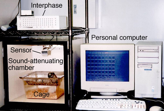
For the quantitative assessment of rat spontaneous motor activity, an activity monitoring system consisting of a sound-attenuating chamber, a sensor monitor and an interface device to convert the activity to digital signals was used [Photo. 1].
The sound-attenuating chamber was an experimental encasement with illumination switched dark/light at 12-hour cycle, extraneous noises cut off and feed and water made freely available. The activities of a rat placed in the chamber were picked up by the sensor mounted on the ceiling, and the sensor signals were converted by the interface, to digital signals representing the number of moves. Signals were analyzed by a personal computer.
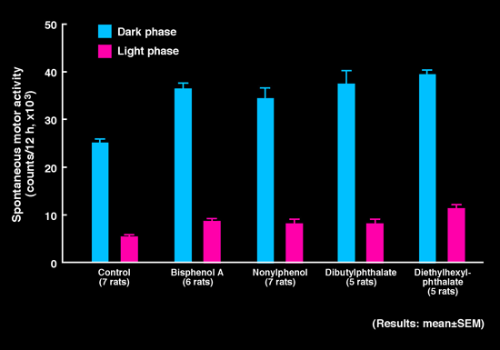
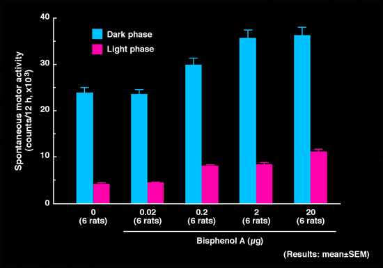
Environmental chemical was administered to the brain (intracisternal administration) of a rat at 5 days of age, and spontaneous motor activity was measured at age 4~5 weeks, which is equivalent to schoolchild stage of human being. As a result, it was found that some of phenols and phthalates augmented spontaneous motor activities similarly to 6-OHDA [Fig. 2]. The significant hyperactivity was observed at a trace dose. For instance, in case of bisphenol A, the injection of 0.2 µg (1 µg = 1/1,000,000 g) caused significant hyperactivity in the rat [Fig. 3].
The histological analysis of the brain revealed that bisphenol A, nonylphenol and p-octylphenol blocked the development of dopaminergic neurons [Photos. 2 and 3].
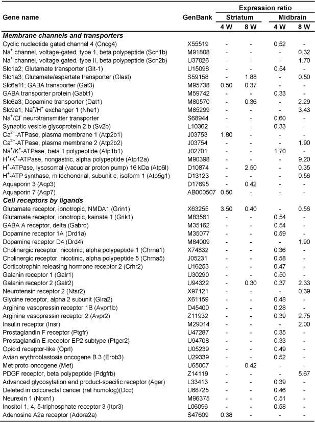
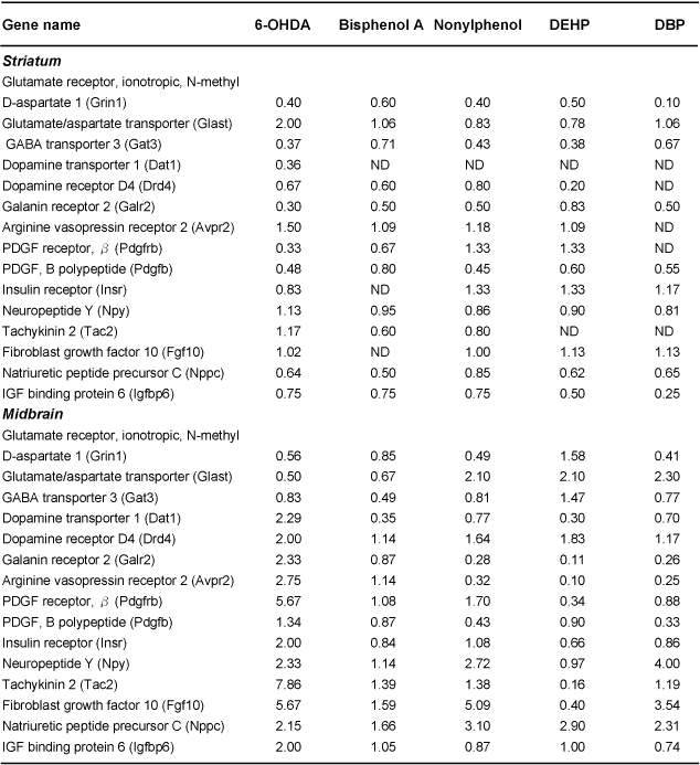
When multiple gene expression was measured in the brain of hyperactivity disorder model rats with 6-OHDA by using DNA chips, the expression of NMDA receptor gene, one of glutamate receptors, was enhanced at the time of hyperactivity onset (4 weeks). Subsequently, at 8 weeks of age, augmented gene expression was recognized for glutamate transporter, dopamine transporter and dopamine D4 receptor [Table 1]. On the other hand, gene expression profiles in hyperactive rats induced by environmental chemicals showed a great variation and differed from those of 6-OHDA [Table 2].
These results suggest that environmental chemicals exert toxic effects not only to dopaminergic neuorns but also to multiple kinds of neurons, such as noradrenergic and serotonergic.
In most of evaluation tests for environmental chemicals, chemicals are administered to pregnant rats and later the effects for offspring rats are examined. However, as the migration of chemicals to the brain of young rats is not fully understood at present, the Research Group is collecting basic data on the effects of direct administration to the brain. The neurotoxicity of environmental chemicals revealed by the screening test not always derives one-track way of conclusion that chemicals in question are responsible to human mental disorders. Further efforts are needed for demonstrating the migration of environmental chemicals to the brain. However, as the behavioral pattern of experimental animals is used as an indicator in this technique, it will be helpful as effective screening for environmental chemicals with neurotoxicity.
 |
|
Photo. 1. A configuration of spontaneous motor activity monitoring system Sound-attenuating chamber |
 |
|
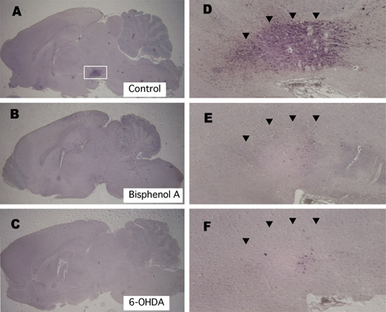
Photo. 2. Tyrosine hydroxylase immunoreactivity after the administration of bisphenol A and 6-hydroxy dopamine. |
Six groups of 5-day-old rats were administered into the brain with solvent (A, D), bisphenol A (B, E) and 6-OHDA (C, F). At 8 weeks of age, the brain was dissected out and stained immunohistochemically by use of anti-tyrosine hydroxylase (TH) antibody. D, E and F represent enlarged views of the midbrain indicated with square in A. In the normal animal administered with solvent only, dopaminergic neurons in the ventral part of the midbrain (substantia nigra and ventral tegmental area) are deeply stained (arrow in D). Bisphenol A lowered the TH immunoreactivity, similarly to 6-OHDA (arrows in E and F). These results indicate that the development of dopaminergic neurons was suppressed.
 |
|
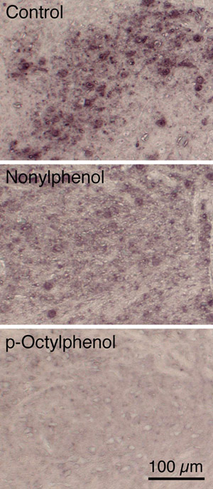
Photo. 3. Tyrosine hydroxylase immunoreactivity after the administration of nonylphenol and p-octylphenol. |
Five-day-old rats were administered into the brain with nonylphenol and p-octylphenol, and at 8 weeks of age, the brain was dissected out and stained immunohistochemically by use of anti-tyrosine hydroxylase (TH) antibody. Pictures show the ventral part of the midbrain. Both chemicals reduced the TH immunoreactivity, suggesting that the development of dopaminergic neurons were inhibited.
 |
|
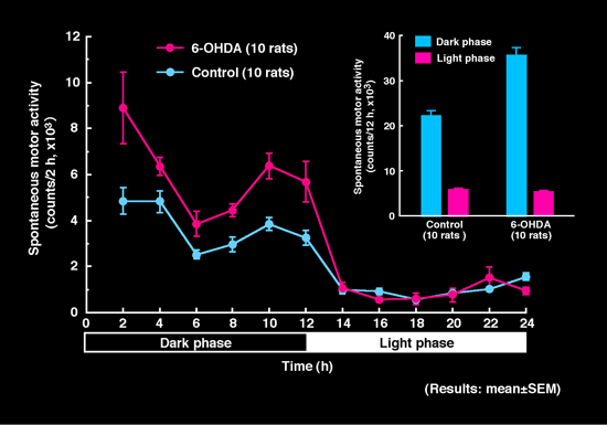
Fig. 1. Effects of 6-hydroxydopamine on spontaneous motor activity in rats. |
Five-day-old rats were administered into the brain with 6-hydroxydopamine (6-OHDA), and at age of 4~5 weeks, the spontaneous motor activity was measured. The measurement was carried out under illumination conditions of dark (12 hrs)/light (12 hrs) cycles. Each point represents motor activity count in every 2-hour. Motor activity in every 12-hour is shown by a bar chart at the top right for comparison. Nocturnal rats showed higher activity in the dark phase than in the light phase. When 6-OHDA was administered, the activity in the dark phase was significantly augmented in comparison to normal animals administered with solvent only. The activity in the light phase presented no difference between the two groups.
 |
|
Fig. 2. Effects of environmental chemicals on spontaneous motor activity in rats. |
Five-day-old rats were administered into the brain with environmental chemicals and at age of 4~5 weeks, the spontaneous motor activity was measured. The measurement was carried out under illumination conditions of dark (12 hrs)/light (12 hrs) cycles. Each column represents motor activity count in every 12-hour. After the administration of environmental chemicals, such as bisphenol A, nonylphenol, dibutylphthalate and diethylhexylphthalate, the activity was significantly augmented both in the dark and light phases, in comparison to normal animals administered with solvent only. The hyperactivity in the light phase that was not recognized in case of the deficit in the development of dopaminergic neurons with 6-OHDA, suggests difficulty in falling asleep in the bright environment.
 |
|
Fig. 3. Effects of bisphenol A on spontaneous motor activity in rats. |
Five-day-old rats were administered into the brain with bisphenol A and at age of 4~5 weeks, the spontaneous motor activity was measured. The measurement was carried out under illumination conditions of dark (12 hrs)/light (12 hrs) cycles. Each column represents motor activity count in every 12-hour. When the dose of bisphenol A was more than 0.2 µg, motor activity was significantly increased both in the dark phase and in the light phase. These results indicate that bisphenol A augments the motor activity in dose-dependent manner.
 |
|
Table 1. Gene expressions affected by 6-OHDA. |
Five-day-old rats were administered with 6-hydroxydopamine and at age of 4 and 8 weeks, the gene expression in the striatum and the midbrain was examined. The results are expressed as an expression ratio in comparison to the expression in normal rats administered with solvent only. The table lists genes with expression ratio greater than 1.69. Unchanged cases are shown by “-“. Abbreviations are based on LocusLink (http://www.ncbi.nih.gov/LocusLink/index.html). Slc, solute carrier family; PDGF, platelet-derived growth factor.
 |
|
Table 2. Changes in gene expression in the striatum and the midbrain of 8-week-old rats. |
Five-day-old rats were administered with chemicals and at age of 8 weeks, the gene expression in the striatum and the midbrain was examined. The results are expressed as an expression ratio in comparison to the expression in normal rats administered with solvent only. “ND” means not detected because of low expression. Abbreviations are based on LocusLink (http://www.ncbi.nih.gov/LocusLink/index.html). DEHP, diethylhexylphthalate; DBP, dibutylphthalate.