Point
A “photon microscope” capable of, even with extremely weak light which is unobservable by conventional optical microscopes, observing a clear color image using a superconducting photodetector has been developed first-ever in the world.
 |
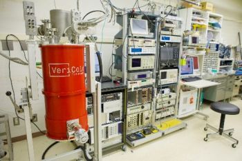 |
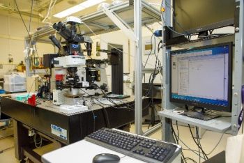
| Sample stage part and color image construction part (left), sensor part and spectrum measurement part (right) of the photon microscope |
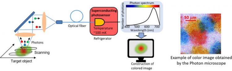
Outline of the photon microscope and example of measured image
|
Background
Optical microscopes are often used for color image observation. However, when light is extremely weak, captured images become dark and unclear. AIST has been developing optical detection technology using superconductivity to detect photons with high accuracy. When one photon is incident on a superconducting photodetector developed by AIST, the energy of the photon temporarily breaks a superconducting state to change electrical resistance, thus making it possible to detect the photon. In addition, from the magnitude of the change in resistance, the wavelength of the photon can also be discriminated.
Outcomes and Methods
We have developed the photon microscope using the superconducting photodetector. Extremely weak light from a target point of a sample is focused and guided to the sensor in a refrigerator (temperature: 100 mK) through an optical fiber. The superconducting photodetector detects photons one by one and measures the wavelengths of them. The color of the target point is discriminated from the number and wavelengths of photons detected within an exposure time. By scanning the sample, a color image is obtained. In the example of measured image (figure), the number of photons from one measurement point was approximately twenty in average (exposure time: 50 ms/point).
Future Plan
We will attempt observing weak emission from living cells stained using multicolor dyes, which has been unobservable, and verify the effectiveness of the photon microscope in the fields of drug discovery and treatment targeting at cells. We will also work on the development of color videography by fabricating a multi-element sensor.
Links
Contact
 |
|
 |
|
Quantum Optical Measurement Group, Research Institute for Physical Measurement
Daiji Fukuda, Leader (left)
Kazuki Niwa, Planning Officer(right)
AIST Tsukuba Central 3, 1-1-1 Umezono, Tsukuba, Ibaraki 305-8563 Japan
|