Takahiro Tanaka (Research Scientist) and Norio Saito (Section Chief), the Ionizing Radiation Section, the Quantum Radiation Division, the Metrology Institute of Japan (Director: Masahiro Okaji) of the National Institute of Advanced Industrial Science and Technology (AIST) (President: Hiroyuki Yoshikawa) have established the X-ray dose standard for the mammography used in the breast cancer screening. Concurrent with this development, a calibration service for the dosemeter in mammography X-ray beam quality has been started from March 5, 2009 (for more details, please visit http://www.nmij.jp/service/P/calibration/).
Mammography uses low-energy X-rays from molybdenum to image breast tissue. The X-ray property (beam quality) of this equipment is different from the property of the X-rays from other X-ray diagnostics such as a chest X-ray system. Therefore, a unique dose standard for accurately measuring the mammography X-ray radiation dose is needed. However, such dose standard has not yet been established in Japan despite the fact that clinical settings have demanded the development of such dose standard for mammography X-ray.
This newly established X-ray dose standard will ensure measurement traceability of radiation dose in the mammography, which can in turn improve the precision of the mammography and meet the needs of the clinical settings. Furthermore, this development ensures the objectivity/reproducibility of mammography diagnosis, which will make Japan internationally competitive in mammography field.
 |
|
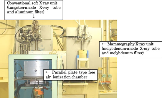
National standard devices for calibrating the mammography dosemeter |
Recently, the importance of early detection and early treatment of breast cancer has been emphasized, and this has resulted in an increase in the number of examinees who underwent X-ray imaging of breasts (mammography) in the breast cancer screening since 2000. The number of mammography examinees has since then increased and exceeded the number of examinees who received only clinical breast examinations in 2005. In 2006, the number reached approximately 1.63 million (Source: MHLW Statistical Database). Since the number of mammography examinees has steadily increased, more attention should be focused on the precision control of the mammography equipment.
In mammography, examinees are exposed to similar amounts of radiation dose that they receive from nature for one year. Therefore, to minimize and precisely manage radiation dose in mammography, the appropriate evaluation of the radiation dose in mammography is critical. Currently, radiation dose has been evaluated in accordance with the national standard for X-ray examinations such as chest X-ray imaging. However, the beam quality of X-ray from a molybdenum tube used in mammography differs from that emitted by a tungsten tube in the typical X-ray systems. Therefore, radiological technologists who are responsible for the precision control in the mammography have waited for the establishment of a new national dose standard suitable for low-energy X-ray used in mammography (mammography X-ray) to more accurately evaluate the dose.
AIST has developed, maintains, and disseminates the dose standard of X-rays using a tungsten tube. The dose standard mainly plays a role in radiation protection and radiography and CT imaging. Recently, the establishment of the dose standard for mammography X-ray using a molybdenum tube has been desired because of an increase in the number of mammography examinees. Therefore, the development of the dose standard for mammography X-ray was started in 2007, and correction factors to define the standard have been determined through experiments and simulations.
Generally, dosemeters for determining X-ray dose are likely to provide different values depending on beam quality even if the same dose is radiated. Therefore, dosemeters should be calibrated based on the beam quality of the X-ray system used in clinical settings. In particular, the system that uses low-energy X-ray, such as in mammography, requires more accurate dosemeter calibration because the sensitivity of the dosemeter varies depending on X-ray energy.
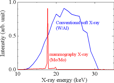
Figure 1 shows the energy distribution of X-rays radiated by a tungsten tube used in the typical X-ray standard and the mammography X-ray. The energy of the X-rays from a tungsten tube is widely distributed, while characteristic X-rays (17.4 keV) are generated from the mammography equipment. Thus, beam quality significantly varies depending on the system so that the dose standard specific to the mammography X-ray is essential.
 |
|
Figure 1. Energy distribution of the mammography X-ray and the X-ray radiated from a tungsten tube obtained by simulation |
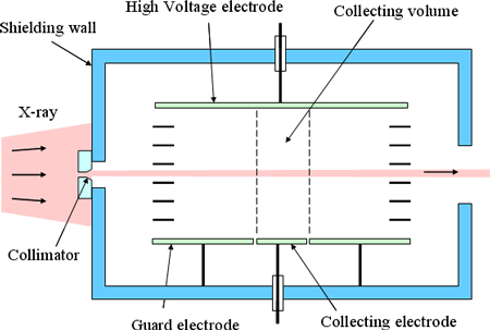
A parallel plate type free air ionization chamber, shown in Fig. 2, was used as the national standard for an absolute measurement of the mammography X-ray dose. X-rays from an X-ray tube pass through a collimator (a small cylindrical hole) to the chamber. The dose was obtained by measuring the charge of ions produced in the chamber. To obtain the accurate X-ray dose, various correction coefficients should be determined either in experiments or by simulation. These coefficients vary depending on the beam quality of the X-ray (e.g., tube voltage) so that the standard was set based on the beam quality.
The calibration coefficients of a dosemeter were determined using the conventional X-ray dose standard and the mammography X-ray dose standard to find any differences. Results are shown in Fig. 3.
 |
|
Figure 2. Schematic diagram of a parallel plate type free air ionization chamber |
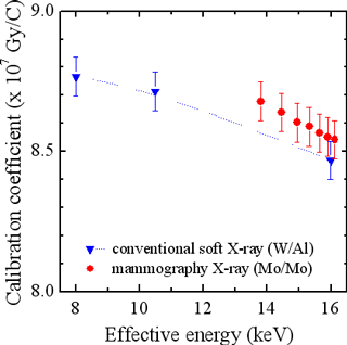 |
Figure 3. Example of a calibration coefficients of a dosemeter
Red: Calibration using the mammography X-ray dose standard
Blue: Calibration using the conventional X-ray dose standard |
As shown in Fig. 3, the calibration coefficients given by the mammography X-ray dose standard was deviated by approximately 1% from that given by the conventional X-ray dose standard. The calibration coefficients varies according to the structure and/or materials of the dosemeters, and the above-mentioned result is not always applicable to other dosemeters. Therefore, the dosemeters should be calibrated using the mammography X-ray dose standard.
This newly developed mammography X-ray dose standard ensures measurement traceability for the evaluation of the mammography X-ray dose at the national level, and it also achieves accurate and reliable dose evaluation of the mammography X-ray. From this time, a more safe and secure breast cancer screening is expected.
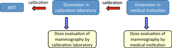
The mammography X-ray dose standard will be subjected to international comparison to obtain international reliability. Furthermore, some mammography X-ray systems use a rhodium tube and filter, which also require the dose standard. We will develop the dose standard for such X-rays and establish the measurement traceability for mammography dose evaluation in the future (Fig. 4).
 |
|
Figure 4. Assumed measurement traceability for the mammography X-ray dose assessment in near future
|