- Moving a step closer to diagnostic applications as a result of the continuous generation of hyperpolarized xenon gas -
The Photonics Research Institute (Director: Masanobu Watanabe) of National Institute of Advanced Industrial Science and Technology (President: Hiroyuki Yoshikawa) (hereinafter referred to as AIST), in collaboration with Toyoko Kagaku Co., Ltd. (President: Hirohisa Kato) (hereinafter referred to as Toyoko Kagaku), has succeeded in building a continuous-flow device that generates hyperpolarized xenon gas with a great efficiency and in developing the device into a commercialized, compact, automated high-performance system for the generation of hyperpolarized xenon gas.
The new device developed by AIST is a result of efforts to increase the level of sophistication of a continuous-flow system for the generation of hyperpolarized xenon gas in nuclear magnetic resonance (NMR) for medical use. In addition to being smaller in size, the device can be connected directly to an NMR apparatus by using simple capillaries. It is expected to be useful in the analysis of pore structures of nanoporous materials, such as those used in fuel cells, and in medical diagnosis technology using sensitive magnetic resonance imaging (MRI) system.
 |
 |
|
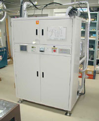
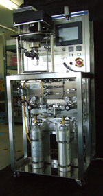
Picture: Left: First continuous-flow type device, Right: Newly developed device (1/5 space occupation) |
The device was exhibited at the Scientific Instruments Show held at the Tokyo Big Sight from Nov. 29 to Dec. 1, 2006.
MRI is a technology that has practical applications in the nondestructive investigation of internal structures of objects in both medicine and industry. For magnetic resonance to occur, an atomic nucleus needs to have magnetic properties that are similar to those of the atomic nucleus of hydrogen. The MRI technique most popularly used in medicine makes use of the atomic nucleus of hydrogen (the proton, 1H) to create images of water present in biomedical tissue and to map the density of hydrogen atomic nuclei in fats: MRI is rarely used in the diagnosis of organs that have low densities of hydrogen nuclei, such as the lungs.
To overcome this problem, researchers have studied the creation of stronger magnetic fields by the use of powerful electromagnets and have attempted to improve detection sensitivity by increasing the efficiency of the detector coils or the detection sequence. Despite these advances, a new sensitization technique based on the principle of NMR had been expected if even higher sensitivities are attainable.
One possible approach involves the use of rare gases in their so-called hyperpolarized state. The rare gases helium, neon, argon, and xenon are stable elements that are unlikely to undergo any chemical reactions and therefore have low toxicities and are safe for living bodies.
It is virtually impossible to observe nuclear magnetic resonance signals from two of the rare gases, 3He and 129Xe, in their normal states because of low sensitivity but, by applying an optical pumping method, it is possible to transform them into a state that produces a strong NMR signal. This state is called the hyperpolarized state.
If the vapor of rubidium (Rb), a low-boiling metal, is irradiated by a special laser beam of circularly polarized light while a magnetic field is applied in parallel (A magnetic field leak of an MRI apparatus is acceptable, but the stronger, the better), the rubidium absorbs the circularly polarized light, resulting in a change in its magnetic properties to a polarized state. This polarized state is transmitted to coexisting 3He and 129Xe raising them into a hyperpolarized state.
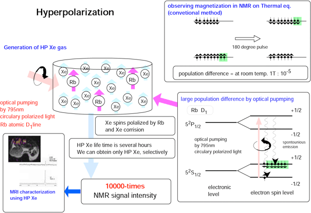
Because the optical pumping method can increase the NMR signal intensities of 3He and 129Xe several tens of thousands of times, it is possible to obtain a hyperpolarized state that gives magnetic resonance signals that are more than 100 times as strong as that of the same volume of water from rare gases that previously could not be used as objects for magnetic resonance because of their low densities (Fig. 1).
Researchers in the United States are developing a device that hyperpolarizes 129Xe at pressures of three or more by using a diode laser with a power of 100 W or more as the source of the excitation radiation and He as the buffer gas. The hyperpolarized 129Xe is trapped by liquid nitrogen and accumulated for high-demand use in MRI for diagnostic applications.
 |
|
Figure 1: Principle of the hyperpolarization and application of NMR/MRI measurements |
When it was the Agency of Industrial and Science Technology, the Ministry of International Trade and Industry, the AIST group, in collaboration with the School of Medicine of Osaka University and University of Occupational and Environmental Health, succeeded in obtaining MRI images of hyperpolarized xenon gas for the first time in Japan, and demonstrated that this gas can be generated at a medical engineering laboratory.
Research continued on the exploitation of the MRI technology in various non-medical applications, such as analyses of the distribution of pore sizes in porous materials with tiny pores, such as fuel cell membranes and catalysts, studies of the dynamic states of gases, and imaging of tiny pores inside refractory bricks for blast furnaces. In a joint research project with Toyoko Kagaku, we developed a practical model that produces hyperpolarized xenon gas at a polarization rate of 2 to 3%. We are accumulating results in several research agencies.
This research was sponsored by the “Research and Development Project to Support Regional Medium- and Small-Sized Companies” promoted by the AIST, and the “Industrial Technology Research Grant Programin 2004” from New Energy and Industrial Technology Development Organization (NEDO) of Japan.
Development of a Continuous-Flow Device for the Generation of Hyperpolarized Xenon Gas
The AIST developed a hyperpolarization device that produces polarized rare gas while it is flowing gas using a flow cell, generates polarized rare gas continuously by installing a nuclear magnetic resonance device at the back, and then performs NMR/MRI measurement without decreasing the polarization rate.
A previous polarization device that used glass cylindrical containers was inefficient because the intensity of the exciting light decreased exponentially with the distance from the entrance face in the direction of the incoming radiation.
The newly developed continuous-flow generation device (Fig. 2) uses a steel flange and a quartz viewport. This device has the following advantages.
-
Unlike the previous glass cell, it allows the internal pressure to be increased to pressure of 10. This permits simultaneous increases in the rate of polarization and the rate of production of the hyperpolarized rare gas.
-
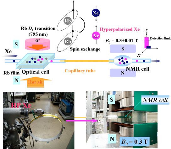
It is possible to install a long capillary to carry the hyperpolarized gas directly to an NMR system (Fig. 3).
-
It is not necessary to position the vacuum apparatus near the NMR system, because rubidium metal is evaporated in a separate vacuum manifold in the newly developed device. Furthermore, the cell can be more easily reused than the previous glass cell.
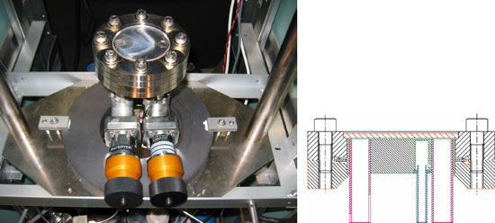 |
Figure 2: Newly developed continuous-flow type hyperpolarized xenon generator
(Left: Picture of the device, Right: Cross section of hyperpolarization cell) |
 |
|
Figure 3: Transportation test of hyperpolarized xenon to NMR equipment using capillary tube. |
In collaboration with the Human Welfare and Medical Engineering Research department, we will attempt to develop a high-speed imaging method for animal use, optimized for hyperpolarized magnetization using an MRI device. We will also continue the research on imaging technology for dynamic analysis within tissues, such as in the analysis of pulmonary function by imaging of porous parts of the lungs by detecting signals from hyperpolarized 129Xe gas, and in measurements of regional cerebral blood flow (rCBF) through temporal changes in the NMR spectrum.